Bacteria
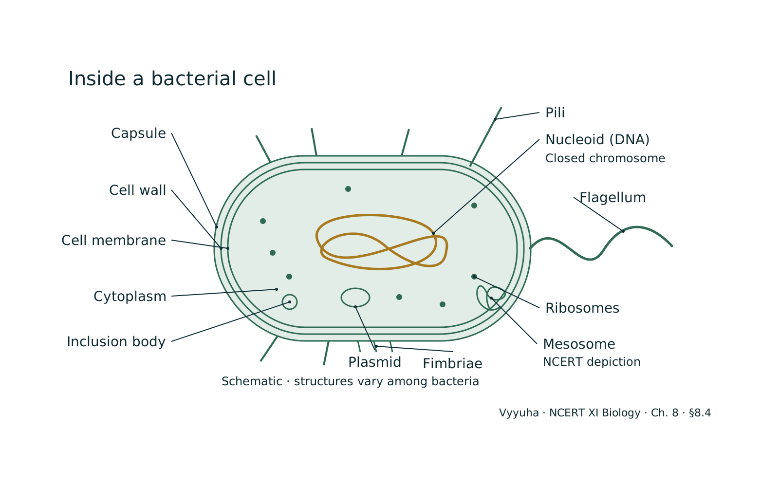
Bacteria are ubiquitous, single-celled prokaryotic microorganisms characterized by the absence of a membrane-bound nucleus and other membrane-bound organelles. Their genetic material, typically a single circular chromosome, resides in the nucleoid region of the cytoplasm. Possessing a rigid cell wall, often composed of peptidoglycan, they exhibit remarkable metabolic diversity, allowing them to th…
Quick Summary
Bacteria are single-celled prokaryotic microorganisms, meaning they lack a membrane-bound nucleus and other membrane-bound organelles. Their genetic material, a single circular chromosome, is located in the nucleoid.
A defining feature is their cell wall, primarily composed of peptidoglycan, which provides structural support and is key to Gram staining classification. They come in various shapes: cocci (spherical), bacilli (rod-shaped), spirilla (spiral), and vibrios (comma-shaped).
Reproduction is mainly by binary fission, a rapid asexual process. Bacteria exhibit immense metabolic diversity, including autotrophs (photo- and chemoautotrophs) and heterotrophs (saprophytes, parasites, symbionts), and can be aerobic or anaerobic.
They play critical roles in ecosystems as decomposers and nutrient cyclers (e.g., nitrogen fixation) and are vital in human health (normal flora, pathogens) and industry (fermentation, biotechnology).
Understanding their structure, metabolism, and classification is fundamental for NEET aspirants.
Full explanation
Bacteria represent one of the most ancient and successful forms of life on Earth, forming the domain Bacteria within the broader classification of life. They are fundamentally prokaryotic organisms, a characteristic that defines much of their cellular architecture and biological processes. Understanding bacteria is crucial not only for microbiology but also for comprehending ecological systems, human health, and biotechnology.
Conceptual Foundation: The Prokaryotic Blueprint
The defining feature of bacteria is their prokaryotic cell organization. Unlike eukaryotic cells, bacterial cells lack a membrane-bound nucleus to enclose their genetic material. Instead, their single, typically circular chromosome is located in a dense region of the cytoplasm called the nucleoid.
They also lack other membrane-bound organelles such as mitochondria, chloroplasts, endoplasmic reticulum, and Golgi apparatus. Despite this structural simplicity, bacterial cells are highly efficient and capable of complex metabolic activities.
Key Structural Components:
- Cell Wall: — A rigid layer outside the plasma membrane, primarily composed of peptidoglycan (also known as murein). This unique polymer, a network of modified sugars (N-acetylglucosamine and N-acetylmuramic acid) cross-linked by short polypeptides, provides structural integrity, protects the cell from osmotic lysis, and determines cell shape. The composition and thickness of the peptidoglycan layer are key to Gram staining, differentiating Gram-positive (thick peptidoglycan, no outer membrane) from Gram-negative (thin peptidoglycan, outer membrane containing lipopolysaccharides).
- Cell Membrane (Plasma Membrane): — A selectively permeable phospholipid bilayer that encloses the cytoplasm. It regulates the transport of nutrients and waste products, houses enzymes for respiration and photosynthesis (in some bacteria), and is involved in cell wall synthesis and DNA replication.
- Cytoplasm: — The jelly-like substance filling the cell, containing water, ions, enzymes, nutrients, and waste products. It is the site of most metabolic reactions.
- Genetic Material: — Primarily a single, circular, double-stranded DNA chromosome located in the nucleoid. Many bacteria also possess smaller, extrachromosomal DNA molecules called plasmids, which carry non-essential but often beneficial genes (e.g., antibiotic resistance, virulence factors).
- Ribosomes: — Responsible for protein synthesis. Bacterial ribosomes are smaller (70S) than eukaryotic ribosomes (80S), a difference exploited by certain antibiotics.
- Flagella: — Long, whip-like appendages used for motility, allowing bacteria to swim towards nutrients or away from toxins (chemotaxis). They are composed of a protein called flagellin and rotate like propellers.
- Pili (Fimbriae): — Shorter, hair-like appendages involved in attachment to surfaces or host cells. Sex pili are specialized for conjugation, the transfer of genetic material between bacteria.
- Capsule/Slime Layer: — An outer layer of polysaccharides or polypeptides, external to the cell wall. A well-organized, tightly attached layer is a capsule, while a diffuse, loosely attached layer is a slime layer. They protect against phagocytosis, desiccation, and aid in adhesion.
- Inclusions: — Storage granules for nutrients like glycogen, poly-beta-hydroxybutyrate, or sulfur.
- Endospores: — Highly resistant, dormant structures formed by some Gram-positive bacteria (e.g., Bacillus, Clostridium) under unfavorable conditions. They can survive extreme heat, radiation, and chemicals, allowing the bacterium to persist for long periods.
Key Principles: Metabolism and Reproduction
Bacteria exhibit an unparalleled range of metabolic strategies:
- Autotrophs: — Produce their own food.
* Photoautotrophs: Use light energy (e.g., cyanobacteria). * Chemoautotrophs: Obtain energy by oxidizing inorganic substances (e.g., nitrifying bacteria, sulfur bacteria).
- Heterotrophs: — Obtain nutrients by consuming organic compounds.
* Saprophytes: Decompose dead organic matter. * Parasites: Live on or in a host, causing harm. * Symbionts: Live in a mutually beneficial relationship with a host.
Respiration:
- Aerobic: — Requires oxygen for cellular respiration.
- Anaerobic: — Does not require oxygen; uses other electron acceptors or fermentation.
- Facultative Anaerobes: — Can grow with or without oxygen.
- Obligate Anaerobes: — Cannot tolerate oxygen.
Reproduction: The primary mode of reproduction is binary fission, an asexual process where a single bacterial cell elongates, duplicates its chromosome, and then divides into two identical daughter cells. This rapid process allows for exponential population growth. Genetic variation can arise through mutations and genetic recombination mechanisms like transformation (uptake of naked DNA), transduction (DNA transfer via bacteriophages), and conjugation (direct transfer via sex pilus).
Classification of Bacteria:
Bacteria are classified based on various criteria:
- Shape:
* Cocci: Spherical (e.g., Staphylococcus, Streptococcus). * Bacilli: Rod-shaped (e.g., Escherichia coli, Bacillus subtilis). * Spirilla: Spiral-shaped, rigid (e.g., Spirillum minor). * Vibrios: Comma-shaped (e.g., Vibrio cholerae). * Spirochetes: Flexible, helical (e.g., Treponema pallidum).
- Gram Staining: — Developed by Hans Christian Gram, this differential staining technique divides bacteria into:
* Gram-positive: Retain crystal violet stain, appearing purple. They have a thick peptidoglycan layer and no outer membrane (e.g., Staphylococcus aureus, Bacillus anthracis). * Gram-negative: Do not retain crystal violet, counterstained pink/red by safranin. They have a thin peptidoglycan layer and an outer membrane (e.g., E. coli, Salmonella typhi).
- Mode of Nutrition: — As discussed above (autotrophs, heterotrophs).
- Oxygen Requirement: — Aerobic, anaerobic, facultative.
Real-World Applications and Significance:
Bacteria are indispensable to life on Earth:
- Ecological Roles:
* Decomposers: Break down dead organic matter, recycling nutrients (e.g., Bacillus, Pseudomonas). * Nitrogen Fixation: Convert atmospheric nitrogen () into ammonia (), making it available to plants (e.g., Rhizobium in legumes, Azotobacter in soil). * Nitrification/Denitrification: Key steps in the nitrogen cycle (e.g., Nitrosomonas, Nitrobacter, Pseudomonas).
- Human Health:
* Normal Flora (Microbiome): Commensal bacteria in the gut, skin, etc., aid digestion, produce vitamins (e.g., Vitamin K), and protect against pathogens (e.g., E. coli in the gut). * Pathogens: Cause diseases (e.g., Mycobacterium tuberculosis causing TB, Salmonella typhi causing typhoid, Clostridium tetani causing tetanus).
- Industrial Applications:
* Food Production: Fermentation of dairy products (yogurt, cheese by Lactobacillus), bread, vinegar. * Antibiotics: Many antibiotics are produced by bacteria (e.g., streptomycin from Streptomyces). * Biotechnology: Used in genetic engineering to produce insulin, vaccines, and other therapeutic proteins (e.g., E. coli). * Bioremediation: Used to clean up oil spills and other pollutants.
Common Misconceptions:
- All bacteria are harmful: — Only a small fraction of bacterial species are pathogenic. Most are harmless, and many are beneficial or essential for life.
- Viruses are bacteria: — Viruses are non-cellular entities, much smaller than bacteria, and require a host cell to replicate. They lack the cellular machinery of bacteria.
- Bacteria are primitive and simple: — While structurally simpler than eukaryotes, bacteria possess highly complex biochemical pathways and sophisticated regulatory mechanisms, allowing them to adapt and thrive in diverse niches.
- Antibiotics kill all bacteria: — Antibiotics are specific. Broad-spectrum antibiotics target a wide range, but narrow-spectrum ones target specific types. Also, antibiotic resistance is a growing problem.
NEET-Specific Angle:
For NEET, focus on the distinguishing features of prokaryotic cells, the unique components like peptidoglycan and plasmids, the different shapes and arrangements, the Gram staining procedure and its implications, various modes of nutrition (especially chemoautotrophs and nitrogen fixers), and the diseases caused by specific bacterial pathogens (e.
g., cholera, typhoid, tetanus, tuberculosis). Understanding the ecological roles, particularly in biogeochemical cycles, is also vital. Questions often test knowledge of specific bacterial examples and their associated functions or diseases.
Key Concepts
Gram staining is a four-step process: 1. Primary stain (crystal violet) stains all cells purple. 2. Mordant…
Nitrogen fixation is the process by which atmospheric nitrogen (), which is unusable by most organisms,…
Bacterial morphology (shape) is a fundamental characteristic used in identification. The four basic shapes…
Often confused with
Side-by-side differences the NEET paper likes to test.
| Aspect | Bacteria | Gram-Positive Bacteria |
|---|---|---|
| Cell Wall Structure | Thick layer of peptidoglycan (20-80 nm), no outer membrane. | Thin layer of peptidoglycan (2-7 nm), presence of an outer membrane containing lipopolysaccharides (LPS). |
| Gram Stain Reaction | Retains crystal violet-iodine complex, appears purple. | Does not retain crystal violet-iodine complex, counterstained pink/red by safranin. |
| Teichoic Acids | Often present in the peptidoglycan layer. | Absent. |
| Periplasmic Space | Generally absent or very small. | Prominent, located between the plasma membrane and outer membrane. |
| Toxins Produced | Primarily exotoxins. | Primarily endotoxins (LPS of outer membrane) and some exotoxins. |
| Sensitivity to Antibiotics | Generally more susceptible to penicillin and lysozyme. | Generally less susceptible to penicillin (due to outer membrane barrier), more susceptible to streptomycin, tetracycline. |
| Examples | *Staphylococcus aureus*, *Streptococcus pneumoniae*, *Bacillus anthracis*, *Clostridium tetani*. | *Escherichia coli*, *Salmonella typhi*, *Pseudomonas aeruginosa*, *Neisseria gonorrhoeae*. |
The fundamental distinction between Gram-positive and Gram-negative bacteria lies in their cell wall architecture, which dictates their response to Gram staining. Gram-positive bacteria possess a thick peptidoglycan layer, allowing them to retain the crystal violet stain and appear purple.
Conversely, Gram-negative bacteria have a thin peptidoglycan layer sandwiched between an inner plasma membrane and an outer membrane containing lipopolysaccharides, causing them to lose the crystal violet and appear pink/red after counterstaining.
This structural difference profoundly impacts their pathogenicity, antibiotic susceptibility, and overall biological characteristics, making Gram staining a cornerstone of bacterial identification and clinical management.
Why it is tested: For NEET, understanding the differences between Gram-positive and Gram-negative bacteria is crucial. Questions frequently test knowledge of their cell wall components, the mechanism of Gram staining, and the implications for antibiotic action. Specific examples of diseases caused by bacteria from each group are also important. This distinction forms a foundational concept in microbiology relevant to human health and disease.
Questions students ask
5 answered on this topic.
What is the primary difference between bacteria and archaea?
While both bacteria and archaea are prokaryotic (lack a membrane-bound nucleus), they differ significantly at the molecular level. Archaea have unique cell wall compositions, often lacking peptidoglycan, and their cell membranes contain branched hydrocarbon chains attached to glycerol by ether linkages, unlike the ester linkages in bacteria.
Their ribosomal RNA sequences are also distinct, placing them in separate domains of life. Archaea are often found in extreme environments, though not exclusively, while bacteria are ubiquitous.
How do bacteria reproduce, and how does this contribute to their rapid evolution?
Bacteria primarily reproduce asexually through binary fission, a process where one cell divides into two identical daughter cells. This rapid division allows for exponential population growth. While binary fission produces clones, genetic variation can arise through spontaneous mutations during DNA replication.
Additionally, bacteria can exchange genetic material horizontally through transformation (uptake of free DNA), transduction (DNA transfer via viruses), and conjugation (direct cell-to-cell transfer via plasmids).
These mechanisms, combined with rapid reproduction, accelerate their adaptation and evolution, including the development of antibiotic resistance.
What is the significance of Gram staining in bacteriology?
Gram staining is a crucial differential staining technique that classifies bacteria into two major groups: Gram-positive and Gram-negative. This distinction is based on differences in their cell wall structure.
Gram-positive bacteria have a thick peptidoglycan layer and retain the crystal violet stain, appearing purple. Gram-negative bacteria have a thin peptidoglycan layer and an outer membrane, preventing crystal violet retention, and are counterstained pink/red by safranin.
This classification is vital for identifying bacteria, guiding antibiotic treatment (as antibiotics often target specific cell wall types), and understanding bacterial pathogenesis.
Are all bacteria harmful to humans?
No, this is a common misconception. While some bacteria are pathogenic and cause diseases, the vast majority of bacterial species are harmless, and many are incredibly beneficial. For instance, the 'normal flora' in our gut aids digestion, produces essential vitamins, and protects against harmful pathogens.
Bacteria are also crucial decomposers in ecosystems, nitrogen fixers in soil, and are widely used in industrial processes like food fermentation and antibiotic production. Only a small percentage of known bacteria are considered human pathogens.
What are endospores, and why are they important?
Endospores are highly resistant, dormant structures produced by certain Gram-positive bacteria, such as Bacillus and Clostridium, when environmental conditions become unfavorable (e.g., nutrient depletion, extreme temperatures).
They are not a form of reproduction but a survival mechanism. Endospores contain the bacterial chromosome and essential cellular components encased in multiple protective layers, making them extraordinarily resistant to heat, radiation, desiccation, and chemical disinfectants.
This allows the bacterium to survive harsh conditions for extended periods and germinate back into a vegetative cell when conditions improve, posing significant challenges in sterilization and food preservation.
Revise in 30 seconds
- Prokaryotic: — No membrane-bound nucleus or organelles.
- Genetic Material: — Single circular (nucleoid), often plasmids.
- Cell Wall: — Peptidoglycan (murein) unique to bacteria.
- Ribosomes: — 70S type.
- Shapes: — Cocci (spherical), Bacilli (rod), Spirilla (spiral), Vibrios (comma).
- Reproduction: — Binary fission (asexual).
- Gram Staining: — Gram-positive (purple, thick peptidoglycan), Gram-negative (pink/red, thin peptidoglycan + outer membrane).
- Nutrition: — Photoautotrophs (e.g., Cyanobacteria), Chemoautotrophs (e.g., Nitrifying bacteria), Heterotrophs (saprophytes, parasites, symbionts).
- Key Roles: — Decomposers, Nitrogen fixers (Rhizobium, Azotobacter), Pathogens.
- Survival: — Endospores (highly resistant dormant structures).
For bacterial shapes, remember: Cute Boys Sing Very Sweetly. Cocci (spherical) Bacilli (rod) Spirilla (rigid spiral) Vibrios (comma) Spirochetes (flexible spiral)