Monocot Root and Stem
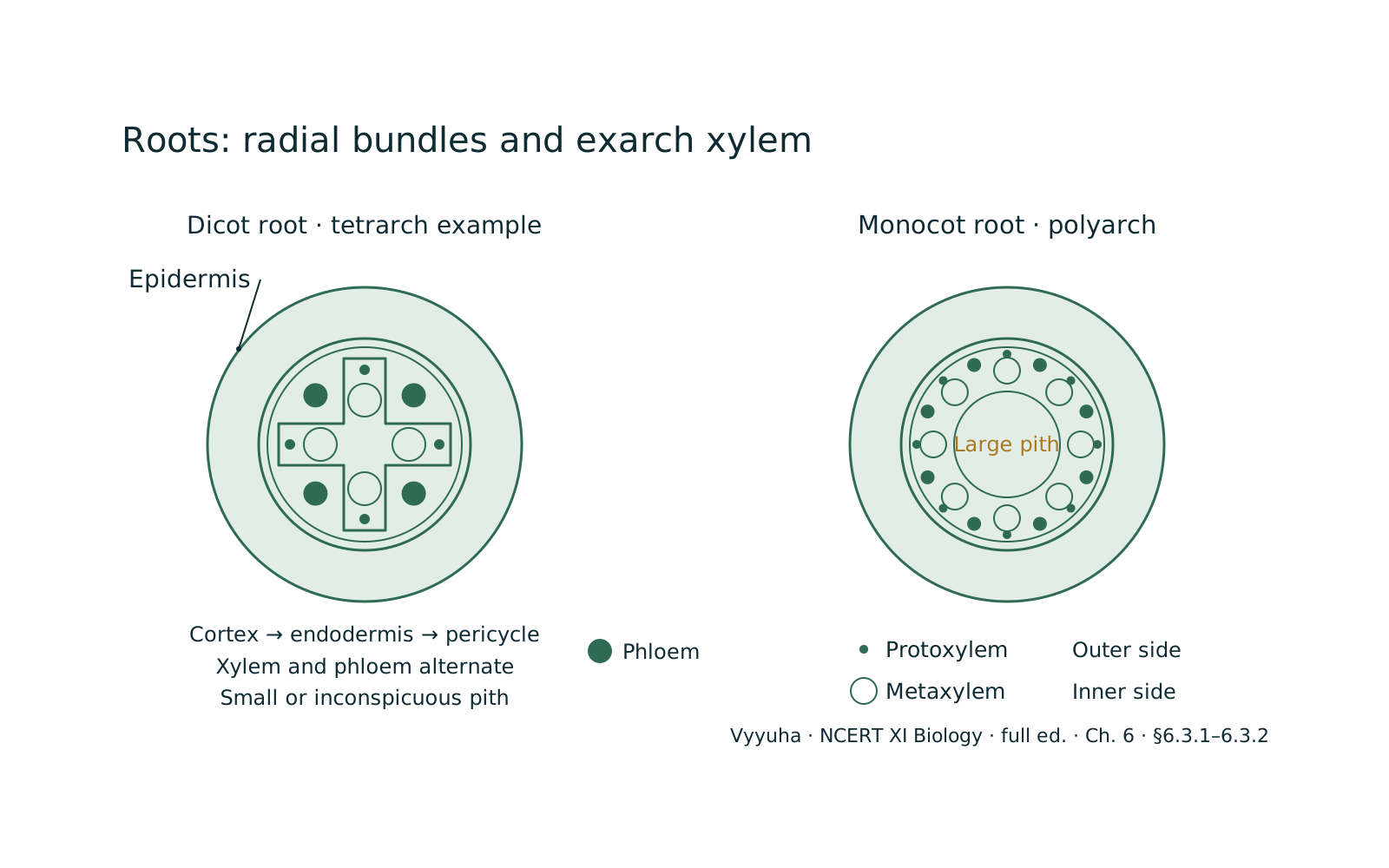
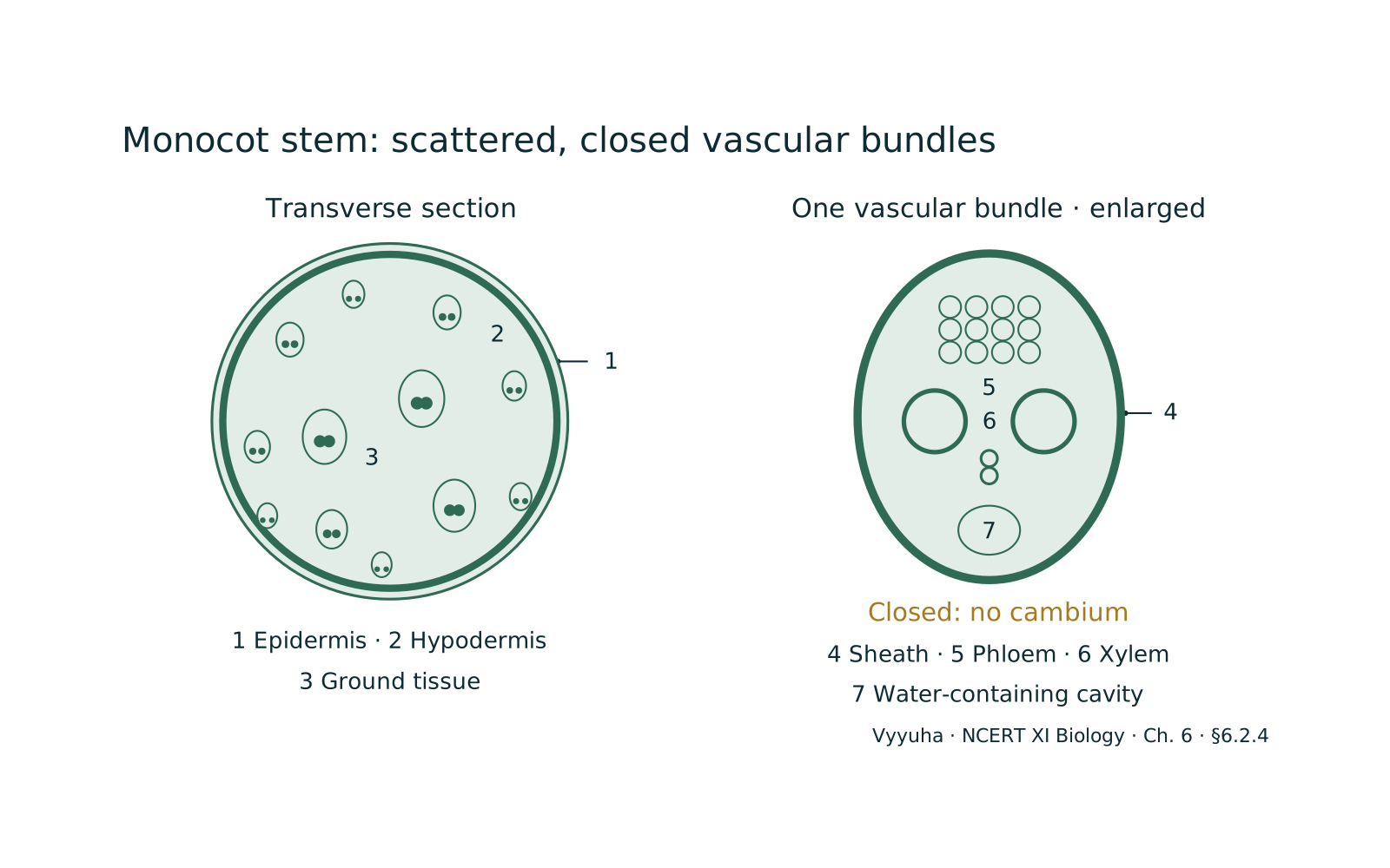
Monocotyledonous plants, or monocots, exhibit distinct anatomical features in their roots and stems that differentiate them from dicotyledonous plants. The monocot root is characterized by a fibrous root system, a polyarch vascular bundle arrangement with numerous xylem and phloem strands, and a prominent pith. In contrast, the monocot stem typically lacks secondary growth, possesses scattered vas…
Quick Summary
Monocot roots are characterized by a fibrous root system, a prominent central pith, and polyarch vascular bundles (many xylem and phloem strands arranged radially). The endodermis features Casparian strips and passage cells.
Lateral roots originate from the pericycle. Monocot stems, on the other hand, exhibit scattered vascular bundles throughout an undifferentiated ground tissue. They possess a sclerenchymatous hypodermis for mechanical support and generally lack secondary growth due to the absence of a vascular cambium (closed vascular bundles).
Each vascular bundle is typically conjoint, collateral, and often surrounded by a sclerenchymatous bundle sheath, with xylem forming a 'V' or 'Y' shape and frequently containing a protoxylem lacuna. These features collectively distinguish monocots from dicots and are crucial for their survival and growth patterns.
Full explanation
The internal organization of monocotyledonous plants, specifically their roots and stems, presents a fascinating study in adaptation and evolutionary divergence from dicots. These anatomical distinctions are not merely academic curiosities but reflect fundamental differences in growth strategies, mechanical support, and resource allocation.
Conceptual Foundation: Plant Tissue Systems
Before delving into the specifics of monocot anatomy, it's essential to recall the three basic tissue systems found in all vascular plants:
- Dermal Tissue System: — The outermost protective layer, primarily the epidermis, which covers the plant body and regulates gas exchange and water loss.
- Ground Tissue System: — Comprises the bulk of the plant body, filling the space between the dermal and vascular tissues. It includes parenchyma, collenchyma, and sclerenchyma, performing functions like photosynthesis, storage, and support.
- Vascular Tissue System: — Responsible for long-distance transport of water, minerals, and sugars throughout the plant. It consists of xylem (water transport) and phloem (food transport).
Monocot roots and stems exhibit unique arrangements and modifications of these tissue systems.
Key Principles: Monocot vs. Dicot Structural Differences
Monocots and dicots diverged early in angiosperm evolution, leading to distinct anatomical blueprints. Key principles guiding monocot anatomy include:
- Absence of Vascular Cambium: — Generally, monocots lack a vascular cambium, which is responsible for secondary growth (increase in girth) in dicots. This means monocot stems typically do not undergo significant secondary thickening.
- Fibrous Root System: — Monocots typically develop a fibrous root system where all roots are more or less equal in size, originating from the stem base, unlike the taproot system of many dicots.
- Parallel Venation: — Leaves typically have parallel veins, influencing the arrangement of vascular tissues in the stem.
- Floral Parts in Multiples of Three: — A general characteristic, though not directly anatomical.
Monocot Root Anatomy
A transverse section (T.S.) of a monocot root reveals a well-organized structure:
- Epidermis (Epiblema): — The outermost single layer of thin-walled, parenchymatous cells. It lacks stomata and cuticle. Some epidermal cells elongate to form unicellular root hairs, which are crucial for water and mineral absorption. This layer is protective.
- Cortex: — Located just beneath the epidermis, the cortex is a broad zone composed of several layers of large, thin-walled parenchymatous cells. These cells are loosely packed, often with intercellular spaces, facilitating gas exchange. The primary function of the cortex is storage of food reserves (e.g., starch) and water.
- Endodermis: — This is the innermost layer of the cortex, consisting of a single ring of barrel-shaped cells. A defining feature is the presence of Casparian strips on their radial and tangential walls. These strips, made of suberin and lignin, are impermeable to water, forcing water and dissolved solutes to pass through the cell membranes (symplastic pathway) rather than between cells (apoplastic pathway). This 'gatekeeper' function ensures selective uptake of substances into the vascular cylinder. Some endodermal cells opposite the protoxylem poles remain thin-walled and lack Casparian strips; these are called passage cells and allow for the passage of water and minerals into the xylem.
- Pericycle: — Situated just internal to the endodermis, the pericycle is a single layer of parenchymatous cells. In monocot roots, the pericycle is responsible for the formation of lateral roots. Unlike dicots, it does not contribute to secondary growth.
- Vascular Bundles: — The vascular tissue forms the central core, known as the stele. In monocot roots, the vascular bundles are radial, meaning xylem and phloem strands are arranged alternately on different radii. A key characteristic is their polyarch nature, meaning there are many (typically more than six, often 8-20 or more) xylem and phloem bundles. The xylem is exarch, with protoxylem (smaller vessels) towards the periphery and metaxylem (larger vessels) towards the center. The phloem bundles are located between the xylem arms.
- Pith: — A prominent and well-developed central region composed of large, thin-walled parenchymatous cells. This is a significant distinguishing feature of monocot roots compared to most dicot roots, where the pith is often small or absent. The pith serves for storage.
Monocot Stem Anatomy
A T.S. of a monocot stem (e.g., maize) reveals a different organization, adapted for upright growth without significant secondary thickening:
- Epidermis: — The outermost protective layer, typically a single layer of parenchymatous cells. It is covered by a thick cuticle to reduce water loss and may bear trichomes (hairs) or stomata.
- Hypodermis: — Immediately below the epidermis, the hypodermis in monocot stems is typically sclerenchymatous. This layer provides significant mechanical support and strength to the stem, especially important in plants that do not undergo secondary thickening to increase girth. It can be several layers thick.
- Ground Tissue: — A hallmark of monocot stems is the undifferentiated ground tissue. Unlike dicot stems, there is no clear distinction into cortex, endodermis, pericycle, and pith. The entire central mass of the stem, internal to the hypodermis, is filled with large, thin-walled parenchymatous cells. These cells are loosely arranged with intercellular spaces and are involved in storage and some metabolic activities.
- Vascular Bundles: — Numerous vascular bundles are scattered throughout the ground tissue, appearing randomly distributed rather than in a ring. This scattered arrangement is a key diagnostic feature. The bundles are typically:
* Conjoint: Xylem and phloem are present together in the same bundle. * Collateral: Phloem is located towards the periphery (outer side) and xylem towards the center (inner side) of the bundle.
* Closed: They lack a vascular cambium between the xylem and phloem, which means they cannot undergo secondary growth. This is why monocot stems generally do not increase much in diameter. * Bundle Sheath: Each vascular bundle is typically surrounded by a sheath of sclerenchymatous cells, providing additional mechanical support and protection.
The vascular bundles are generally smaller and more numerous towards the periphery and larger towards the center of the stem. Within each bundle: * Phloem: Consists of sieve tube elements, companion cells, and phloem parenchyma.
Phloem fibers are generally absent. * Xylem: Typically 'V' or 'Y' shaped. It contains two large metaxylem vessels towards the periphery and one or two smaller protoxylem vessels towards the center.
Often, the lowest protoxylem vessel disintegrates, forming a protoxylem lacuna (a water-filled cavity), which is a characteristic feature of many monocot stems.
Real-World Applications
Understanding monocot root and stem anatomy has practical implications:
- Agriculture: — Knowledge of root systems helps in optimizing irrigation and nutrient delivery for monocot crops like rice, wheat, and corn. Understanding stem structure informs strategies for lodging resistance (preventing stems from falling over) in cereals.
- Plant Identification: — Anatomical features are crucial for identifying plant species, especially in forensic botany or archaeological studies.
- Bioengineering: — Researchers can use this knowledge to genetically modify plants for improved strength, drought resistance, or nutrient uptake.
- Ecological Studies: — Different root and stem architectures are adaptations to various environments, influencing how plants compete for resources and withstand environmental stresses.
Common Misconceptions
- Secondary Growth in Monocots: — A common misconception is that monocots never exhibit any form of secondary growth. While they generally lack a vascular cambium for true secondary thickening, some monocots (e.g., palms, Dracaena, Yucca) show anomalous secondary growth, where a meristematic ring forms in the ground tissue, producing additional vascular bundles and parenchyma, leading to an increase in girth. However, this is not homologous to the dicot vascular cambium.
- Pith in Monocot Stems: — Students often confuse the undifferentiated ground tissue of monocot stems with the distinct pith of dicot stems. The monocot stem lacks a clearly demarcated pith; the entire central region is ground tissue.
- Scattered Bundles Mean Disorganized: — While 'scattered' implies a lack of ring arrangement, the distribution is not entirely random. Smaller bundles are often peripheral, and larger ones central, reflecting an organized pattern for efficient transport and support.
NEET-Specific Angle
For NEET aspirants, the focus should be on distinguishing features. Be prepared to:
- Identify diagrams: — Recognize T.S. of monocot root and stem based on key features (polyarch xylem, prominent pith in root; scattered bundles, sclerenchymatous hypodermis, undifferentiated ground tissue, protoxylem lacuna in stem).
- Recall specific terms: — Casparian strips, passage cells, polyarch, exarch, endarch, conjoint, collateral, closed, bundle sheath, protoxylem lacuna, sclerenchymatous hypodermis.
- Compare and contrast: — Be able to articulate the differences between monocot root and stem, and also between monocot and dicot roots/stems. Questions often test these comparative aspects.
- Functional significance: — Understand why these structures exist (e.g., Casparian strips for selective absorption, sclerenchymatous hypodermis for support).
- Exceptions: — Be aware of exceptions like anomalous secondary growth in some monocots, as these can be tricky questions.
Key Concepts
In a monocot root, the vascular tissue is organized radially, meaning the xylem and phloem strands are…
The arrangement of vascular bundles in a monocot stem is a key distinguishing feature. Unlike the ring-like…
The monocot stem has specific adaptations for mechanical support and water transport. The 'hypodermis,'…
Often confused with
Side-by-side differences the NEET paper likes to test.
| Aspect | Monocot Root and Stem | Monocot Stem |
|---|---|---|
| Overall Structure | Monocot Root: Cylindrical, generally uniform diameter, fibrous root system. | Monocot Stem: Cylindrical, often tapering, generally upright, no significant secondary thickening. |
| Epidermis | Monocot Root: Epiblema, usually with unicellular root hairs, no cuticle, no stomata. | Monocot Stem: Single layer, covered by thick cuticle, may have stomata and trichomes. |
| Hypodermis | Monocot Root: Absent. | Monocot Stem: Present, typically sclerenchymatous, providing mechanical support. |
| Ground Tissue | Monocot Root: Differentiated into cortex, endodermis, and pericycle. Cortex is broad, parenchymatous. | Monocot Stem: Undifferentiated, continuous mass of parenchymatous cells, no distinct cortex, endodermis, pericycle, or pith. |
| Vascular Bundles Arrangement | Monocot Root: Radial, polyarch (many xylem and phloem bundles arranged alternately on different radii). | Monocot Stem: Scattered throughout the ground tissue, not arranged in a ring. |
| Vascular Bundles Type | Monocot Root: Radial, exarch xylem (protoxylem towards periphery, metaxylem towards center). | Monocot Stem: Conjoint, collateral, closed (no cambium), endarch xylem (protoxylem towards center, metaxylem towards periphery). |
| Pith | Monocot Root: Large, well-developed, parenchymatous, central. | Monocot Stem: Absent or very small, as the ground tissue is undifferentiated. |
| Secondary Growth | Monocot Root: Absent. | Monocot Stem: Generally absent (closed vascular bundles), though some show anomalous secondary growth. |
| Protoxylem Lacuna | Monocot Root: Absent. | Monocot Stem: Often present in vascular bundles, formed by disintegration of protoxylem. |
Monocot roots are primarily absorptive and anchoring organs, characterized by a polyarch, radial vascular system with a prominent central pith. Their epidermis forms root hairs, and the endodermis has Casparian strips and passage cells for selective absorption.
In contrast, monocot stems are designed for support and transport, featuring scattered, closed vascular bundles within an undifferentiated ground tissue. A sclerenchymatous hypodermis provides mechanical strength, compensating for the general absence of secondary growth.
The presence of a protoxylem lacuna is also a common distinguishing feature in monocot stems.
Why it is tested: For NEET, understanding these differences is crucial for identifying plant parts from diagrams, answering comparative questions, and recalling specific anatomical terms. Questions frequently test the unique features of monocot roots (polyarch, pith) versus monocot stems (scattered bundles, sclerenchymatous hypodermis, protoxylem lacuna).
Questions students ask
5 answered on this topic.
What is the primary difference in vascular bundle arrangement between monocot and dicot roots?
The primary difference lies in the number and arrangement of xylem and phloem bundles. Monocot roots exhibit a polyarch condition, meaning they have many (typically more than six) xylem and phloem bundles arranged alternately in a ring.
In contrast, dicot roots usually have a tetrarch, triarch, or diarch condition, with fewer (two to six) xylem and phloem bundles, also arranged radially. Monocot roots also typically possess a large, well-developed pith, which is often small or absent in dicot roots.
Why do monocot stems generally not undergo secondary growth?
Monocot stems generally do not undergo secondary growth because their vascular bundles are 'closed,' meaning they lack a vascular cambium. The vascular cambium is a lateral meristem responsible for producing secondary xylem and secondary phloem, leading to an increase in the girth of the stem. Without this cambium, monocot stems cannot form new vascular tissues in the same way dicots do, thus limiting their secondary thickening.
What is the significance of the sclerenchymatous hypodermis in monocot stems?
The sclerenchymatous hypodermis in monocot stems provides crucial mechanical support and strength. Since monocot stems typically lack secondary growth and therefore do not develop extensive woody tissues for support, this tough, lignified layer of sclerenchyma cells acts as a primary structural reinforcement. It helps the stem withstand bending and other physical stresses, allowing the plant to grow upright.
Explain the term 'protoxylem lacuna' in monocot stems.
A protoxylem lacuna is a water-filled cavity often found in the vascular bundles of monocot stems. It forms when the innermost protoxylem vessels (the first-formed xylem elements) disintegrate during the stem's development. This creates a small, empty space within the vascular bundle, which is a characteristic anatomical feature used to identify monocot stems. It is essentially a remnant of the early xylem that has broken down.
How do passage cells in the monocot root endodermis function?
Passage cells are specific endodermal cells in the monocot root that remain thin-walled and lack Casparian strips, unlike the surrounding endodermal cells. They are typically located opposite the protoxylem poles. Their function is to allow water and dissolved minerals to bypass the Casparian strips and enter the vascular cylinder (stele) via the symplastic pathway (through the cell cytoplasm), facilitating the transport of essential substances to the xylem for upward movement.
Revise in 30 seconds
- Monocot Root: — Fibrous root system, polyarch vascular bundles ( xylem/phloem), radial arrangement, exarch xylem, large central pith, endodermis with Casparian strips & passage cells, pericycle for lateral roots.
- Monocot Stem: — Scattered vascular bundles, undifferentiated ground tissue, sclerenchymatous hypodermis, closed vascular bundles (no cambium), conjoint & collateral, endarch xylem (often 'Y' or 'V' shaped), protoxylem lacuna often present, sclerenchymatous bundle sheath.
For Monocot Stem features, remember: Scattered Hypodermis Undifferentiated Closed Protoxylem.
- Scattered vascular bundles
- Hypodermis (sclerenchymatous)
- Undifferentiated ground tissue
- Closed vascular bundles (no cambium)
- Protoxylem lacuna often present