Anatomy of Flowering Plants
- 1TissuesMeristematic Tissues · Permanent TissuesHigh yield
- 2Tissue SystemsEpidermal Tissue System · Ground and Vascular Tissue SystemsHigh yield
- 3Anatomy of Dicot and Monocot PlantsDicot Root and Stem · Monocot Root and StemHigh yield
- 4Secondary GrowthFormation of Secondary Tissues · Annual RingsHigh yield
Plant anatomy is the branch of botany concerned with the internal structure of plants. It delves into the organization of cells into tissues, and tissues into organs, providing a microscopic view of how plants are constructed to perform their various physiological functions. This field is crucial for understanding the adaptations of plants to different environments, their growth patterns, and how …
Quick Summary
Plant anatomy is the study of the internal structure of plants, revealing how cells are organized into tissues and tissues into organs. The plant body is built from three fundamental tissue systems: the epidermal tissue system for protection, the ground tissue system for bulk functions like storage and photosynthesis, and the vascular tissue system for transport (xylem for water, phloem for food).
Growth occurs from meristematic tissues: apical meristems increase length (primary growth), while lateral meristems (vascular cambium, cork cambium) increase girth (secondary growth). Permanent tissues, derived from meristems, are specialized for specific roles and include simple tissues (parenchyma, collenchyma, sclerenchyma) and complex tissues (xylem, phloem).
Key anatomical differences exist between monocots and dicots in their roots, stems, and leaves, particularly in the arrangement of vascular bundles and the presence/absence of secondary growth. Understanding these internal structures is vital for comprehending plant function and adaptation.
Full explanation
The study of plant anatomy provides a fundamental understanding of how plants are structured internally to carry out their life processes. It bridges the gap between cellular biology and whole-plant physiology, revealing the intricate organization that enables plants to thrive.
Conceptual Foundation: The Plant Body Organization
At its most basic, a plant body is composed of cells. These cells, specialized for particular functions, aggregate to form tissues. Tissues, in turn, are organized into tissue systems, and these systems collectively form the various organs of the plant (roots, stems, leaves, flowers). This hierarchical organization ensures efficiency and division of labor within the plant.
Key Principles: Tissue Systems
Flowering plants exhibit three major tissue systems, each with distinct functions and characteristic cellular compositions:
- The Epidermal Tissue System: — This is the outermost protective layer of the plant body, covering roots, stems, and leaves. It comprises epidermal cells, stomata (in leaves and young stems), and epidermal appendages like trichomes (hairs) and root hairs. Its primary functions include protection against mechanical injury, pathogen invasion, excessive water loss, and facilitating gas exchange.
- The Ground Tissue System: — This system constitutes the bulk of the plant body, filling the regions between the epidermal and vascular tissues. It is primarily composed of parenchyma, collenchyma, and sclerenchyma cells. The ground tissue performs various functions depending on its location, including photosynthesis (in leaves), storage (in roots and stems), and support.
- The Vascular Tissue System: — This is the plant's transport system, responsible for the conduction of water, minerals, and organic nutrients throughout the plant. It consists of complex tissues: xylem and phloem. Xylem transports water and dissolved minerals from roots to other parts, while phloem transports synthesized food (sugars) from leaves to storage organs and growing regions.
Meristematic Tissues: The Architects of Growth
Plant growth is indeterminate, meaning they can grow throughout their lifespan, thanks to specialized regions of actively dividing cells called meristems. These are classified based on their location:
- Apical Meristems: — Located at the tips of roots and shoots, responsible for primary growth (increase in length). The shoot apical meristem produces the stem and leaves, while the root apical meristem produces the root cap and the main root body.
- Intercalary Meristems: — Found at the base of internodes or leaf blades, particularly in grasses. They contribute to the elongation of organs and are responsible for the regrowth of parts removed by grazing herbivores.
- Lateral Meristems: — Responsible for secondary growth (increase in girth or thickness). These include the vascular cambium (producing secondary xylem and phloem) and the cork cambium (producing periderm, the outer bark).
Permanent Tissues: Specialized for Function
Cells derived from meristems differentiate and mature to form permanent tissues, which have lost the ability to divide. They are categorized into simple and complex types:
- Simple Permanent Tissues: — Composed of only one type of cell.
* Parenchyma: Most abundant, living cells with thin walls, involved in storage, photosynthesis, and secretion. Found in cortex, pith, and mesophyll. * Collenchyma: Living cells with unevenly thickened cell walls, providing mechanical support to young stems and petioles.
Found beneath the epidermis in dicot stems. * Sclerenchyma: Dead cells with thick, lignified walls, providing mechanical support and protection. Includes fibers (elongated) and sclereids (variously shaped, often stone cells).
- Complex Permanent Tissues: — Composed of more than one type of cell, working together as a unit.
* Xylem: Consists of tracheids, vessels, xylem parenchyma, and xylem fibers. Tracheids and vessels are the main water-conducting elements. Xylem also provides mechanical support. * Phloem: Consists of sieve tube elements, companion cells, phloem parenchyma, and phloem fibers. Sieve tube elements conduct food, while companion cells regulate their activity.
Anatomy of Dicotyledonous and Monocotyledonous Plants
Understanding the internal structure of roots, stems, and leaves is critical, especially the differences between dicots and monocots.
- Dicot Root: — Possesses a central stele with a limited number of radial vascular bundles (2-6 xylem arms), a well-developed pith is often absent or very small. The cortex is broad, and secondary growth occurs.
- Monocot Root: — Has a larger number of radial vascular bundles (more than 6 xylem arms), a prominent pith, and generally does not undergo secondary growth.
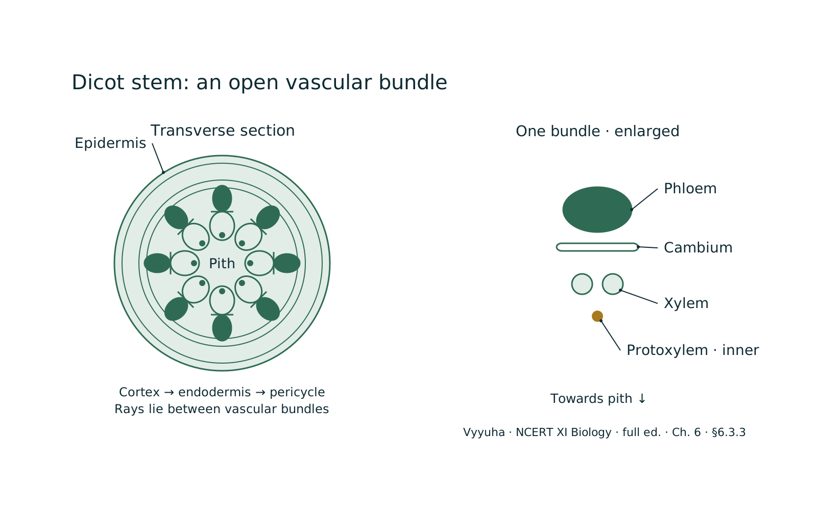
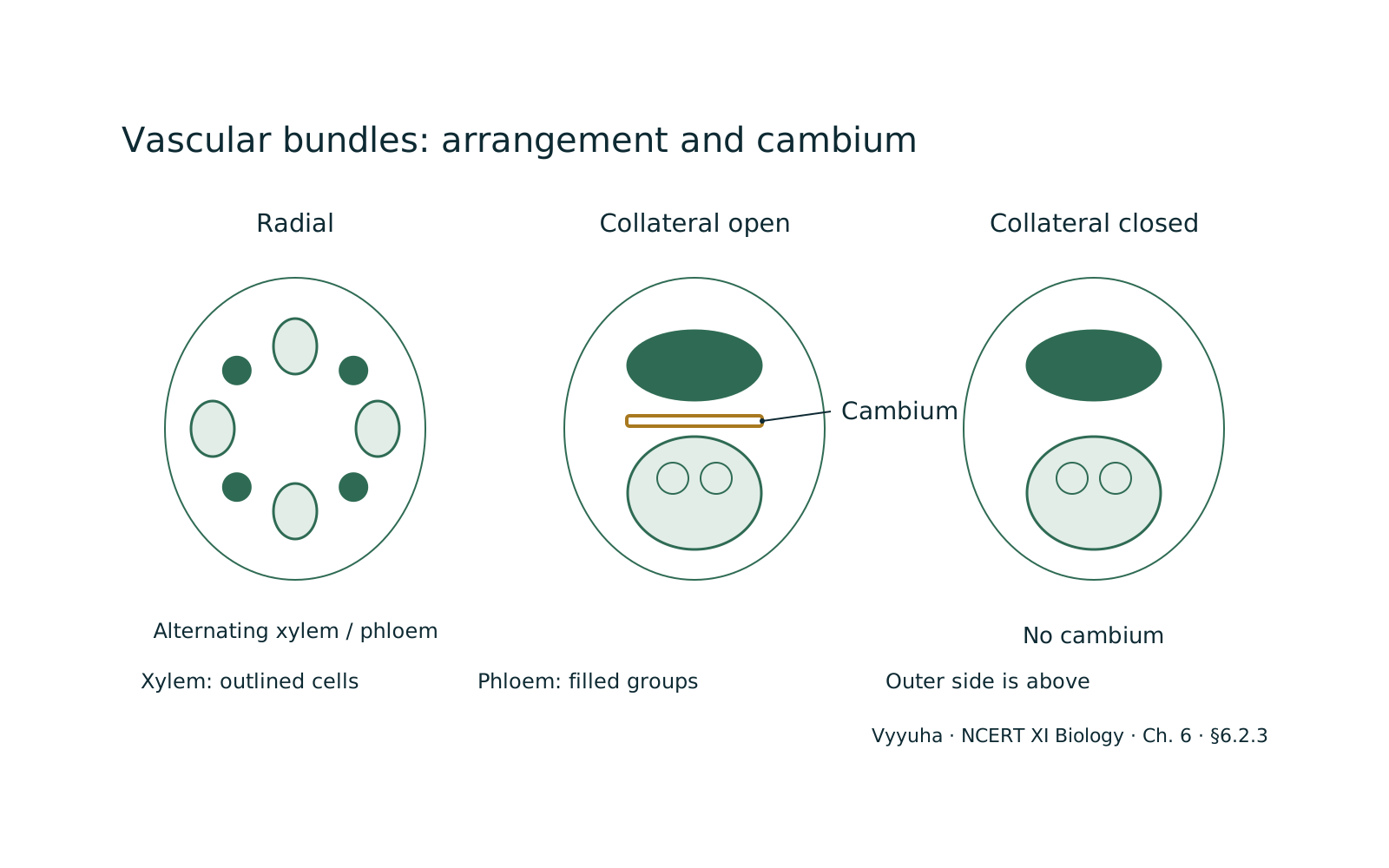
- Dicot Stem: — Vascular bundles are arranged in a ring, open (possessing cambium), and conjoint. A distinct pith is present in the center, and a well-differentiated cortex and endodermis are observed. Secondary growth is common.
- Monocot Stem: — Vascular bundles are scattered, closed (lacking cambium), and conjoint. Ground tissue is undifferentiated, and a distinct pith is absent. Secondary growth is typically absent, though some monocots show anomalous secondary thickening.
- Dicot Leaf (Dorsiventral Leaf): — Exhibits distinct upper (adaxial) and lower (abaxial) epidermises. The mesophyll is differentiated into palisade parenchyma (elongated, tightly packed cells for photosynthesis) and spongy parenchyma (irregularly shaped cells with air spaces for gas exchange). Stomata are more numerous on the lower epidermis. Vascular bundles are surrounded by bundle sheath cells.
- Monocot Leaf (Isobilateral Leaf): — Mesophyll is not differentiated into palisade and spongy parenchyma; cells are more or less uniform. Stomata are equally distributed on both epidermises. Some grass leaves have bulliform cells (large, empty, colourless cells) on the adaxial epidermis, which help in rolling the leaf to reduce water loss.
Secondary Growth: Increasing Girth
Secondary growth, primarily observed in dicot stems and roots, involves an increase in the diameter of the plant body. This process is facilitated by two lateral meristems:
- Vascular Cambium: — Forms a complete ring between the xylem and phloem. It cuts off secondary xylem towards the inside and secondary phloem towards the outside, leading to an increase in the girth of the stem/root. The activity of the vascular cambium is influenced by seasonal variations, leading to the formation of annual rings (spring wood and autumn wood).
- Cork Cambium (Phellogen): — Develops in the cortex region. It cuts off cork (phellem) towards the outside and secondary cortex (phelloderm) towards the inside. The phellem, phellogen, and phelloderm together constitute the periderm, which replaces the epidermis as the protective outer layer in older stems and roots.
Real-world Applications:
Understanding plant anatomy has numerous practical applications. In agriculture, knowledge of vascular tissue arrangement helps in grafting techniques. Identifying wood types based on anatomical features is crucial in forestry and timber industries.
Plant breeders use anatomical insights to select for desirable traits, such as stronger stems (due to more sclerenchyma) or more efficient water transport. Forensic botany can use plant anatomical features to identify plant fragments at crime scenes.
Moreover, the study of plant anatomy is fundamental to understanding plant diseases and developing resistant varieties.
Common Misconceptions:
- Morphology vs. Anatomy: — Students often confuse these. Morphology is external appearance; anatomy is internal structure. A plant's external shape (morphology) is often a reflection of its internal organization (anatomy).
- Primary vs. Secondary Growth: — Primary growth is increase in length (apical meristems); secondary growth is increase in girth (lateral meristems). Not all plants undergo significant secondary growth (e.g., most monocots).
- Monocot vs. Dicot: — While there are general rules, some monocots show anomalous secondary growth, and some dicots may have features resembling monocots. It's important to understand the typical distinguishing features rather than rigid classifications.
- Function of Xylem and Phloem: — While xylem primarily transports water and minerals and phloem transports food, both also provide mechanical support due to the presence of fibers.
NEET-Specific Angle:
For NEET, the focus is heavily on comparative anatomy of monocot and dicot roots, stems, and leaves. Students must be able to identify different tissues and their arrangement from diagrams. Questions frequently test the functions of specific cells and tissues (e.
g., companion cells, bulliform cells, tracheids). Understanding the process and products of secondary growth, including the formation of annual rings, heartwood, and sapwood, is also crucial. Memorizing the location and characteristics of different meristems and permanent tissues is key.
Diagrams are often provided, and students are asked to identify labeled parts or infer properties based on the visual representation.
Key Concepts
Vascular bundles are the plant's transport highways, containing xylem and phloem. Their arrangement and…
Meristems are regions of continuous cell division, producing new cells. These newly formed cells then undergo…
Leaves exhibit specific anatomical features adapted for photosynthesis and water regulation. The mesophyll…
Often confused with
Side-by-side differences the NEET paper likes to test.
| Aspect | Anatomy of Flowering Plants | Monocot vs. Dicot Stem Anatomy |
|---|---|---|
| Vascular Bundle Arrangement | Dicot Stem: Arranged in a ring | Monocot Stem: Scattered throughout the ground tissue |
| Vascular Bundle Type | Dicot Stem: Conjoint, collateral, and open (cambium present) | Monocot Stem: Conjoint, collateral, and closed (cambium absent) |
| Ground Tissue | Dicot Stem: Differentiated into cortex, endodermis, pericycle, and pith | Monocot Stem: Undifferentiated ground tissue |
| Secondary Growth | Dicot Stem: Undergoes secondary growth (increase in girth) | Monocot Stem: Generally does not undergo secondary growth |
| Bundle Sheath | Dicot Stem: Usually absent or parenchymatous | Monocot Stem: Prominent sclerenchymatous bundle sheath around vascular bundles |
The anatomical differences between monocot and dicot stems are fundamental for plant identification and understanding their growth patterns. Dicot stems exhibit a highly organized internal structure with vascular bundles in a ring and the capacity for secondary thickening, allowing them to form woody structures.
Monocot stems, conversely, have a more uniform ground tissue and scattered vascular bundles, which are typically 'closed,' limiting their ability to increase in girth. These distinctions reflect different evolutionary strategies and adaptations.
Why it is tested: For NEET, distinguishing between monocot and dicot anatomy is a frequently tested concept. Questions often involve identifying diagrams or listing characteristic features. Understanding these differences is crucial for correctly interpreting plant structures and their functions, which forms a significant part of the Biology syllabus.
Questions students ask
6 answered on this topic.
What is the primary difference between primary and secondary growth in plants?
Primary growth refers to the increase in the length of the plant, occurring at the tips of roots and shoots due to the activity of apical meristems. It results in the formation of primary tissues. Secondary growth, on the other hand, is the increase in the girth or thickness of the plant, primarily in dicots and gymnosperms. It is facilitated by lateral meristems, namely the vascular cambium and cork cambium, leading to the formation of secondary xylem, secondary phloem, and periderm.
How can I distinguish between a monocot stem and a dicot stem based on their anatomy?
Several key features differentiate them. A dicot stem typically has vascular bundles arranged in a ring, and these bundles are 'open' (contain cambium), allowing for secondary growth. It also has a distinct pith and cortex.
In contrast, a monocot stem has scattered vascular bundles that are 'closed' (lack cambium), preventing secondary growth. The ground tissue is undifferentiated, and a distinct pith is usually absent. The presence of a sclerenchymatous bundle sheath around vascular bundles is also characteristic of monocot stems.
What are annual rings and what is their significance?
Annual rings, also known as growth rings, are concentric circles visible in the cross-section of a woody stem. They are formed due to the differential activity of the vascular cambium throughout the year.
During spring, the cambium is more active, producing wider vessels (spring wood or early wood). In winter, activity decreases, forming narrower vessels (autumn wood or late wood). Each ring represents one year's growth.
Their significance lies in determining the age of a tree (dendrochronology) and providing insights into past climatic conditions.
What is the function of the periderm and how is it formed?
The periderm is a protective tissue that replaces the epidermis in older stems and roots, especially in plants undergoing secondary growth. It provides protection against water loss, mechanical injury, and pathogen invasion.
The periderm is formed by the activity of the cork cambium (phellogen), a lateral meristem that develops in the cortex. The phellogen cuts off cork cells (phellem) towards the outside and secondary cortex cells (phelloderm) towards the inside.
Together, phellem, phellogen, and phelloderm constitute the periderm.
Explain the role of companion cells in the phloem.
Companion cells are specialized parenchyma cells closely associated with sieve tube elements in the phloem. They are living cells with a prominent nucleus and cytoplasm, unlike the enucleated sieve tube elements.
Their primary role is to assist the sieve tube elements in their function of food conduction. They load sugars into the sieve tube elements and unload them, providing metabolic support and regulating the activity of the sieve tube elements through plasmodesmatal connections.
This symbiotic relationship is crucial for efficient translocation of nutrients.
What are bulliform cells and where are they found?
Bulliform cells are large, empty, colourless cells found in the adaxial (upper) epidermis of many monocot leaves, particularly grasses. These cells play a crucial role in reducing water loss. When the plant experiences water stress, bulliform cells lose turgor, become flaccid, and cause the leaf blade to roll inwards. This rolling reduces the exposed surface area, thereby minimizing transpiration and conserving water. Upon rehydration, these cells regain turgor, and the leaf unrolls.
Revise in 30 seconds
- Meristems: — Apical (length), Intercalary (length, grasses), Lateral (girth - vascular cambium, cork cambium).
- Simple Permanent Tissues:
- Parenchyma: Living, thin-walled, storage, photosynthesis. - Collenchyma: Living, unevenly thickened walls, flexible support (young parts). - Sclerenchyma: Dead, thick lignified walls, rigid support (mature parts).
- Complex Permanent Tissues:
- Xylem: Water/minerals transport. Components: Tracheids, Vessels, Xylem Parenchyma, Xylem Fibers. - Phloem: Food transport. Components: Sieve Tube Elements, Companion Cells, Phloem Parenchyma, Phloem Fibers.
- Tissue Systems: — Epidermal (protection), Ground (bulk, storage, photo), Vascular (transport).
- Monocot vs. Dicot: — Key differences in root, stem, leaf anatomy (vascular bundle arrangement, pith, cambium, mesophyll).
- Secondary Growth: — Vascular cambium (secondary xylem/phloem, annual rings), Cork cambium (periderm: phellem, phellogen, phelloderm).
To remember the components of Xylem and Phloem: Xylophone Tunes Very Pleasantly For X-mas (Xylem: Tracheids, Vessels, Parenchyma, Fibers) People Sing Carols Proudly For Presents (Phloem: Sieve tubes, Companion cells, Parenchyma, Fibers)