Anatomy of Dicot and Monocot Plants
Plant anatomy is the study of the internal structure of plants. Understanding the anatomical differences between dicotyledonous (dicot) and monocotyledonous (monocot) plants is fundamental to comprehending their diverse physiological functions, growth patterns, and evolutionary adaptations. These differences are evident in their roots, stems, and leaves, reflecting distinct strategies for water an…
Quick Summary
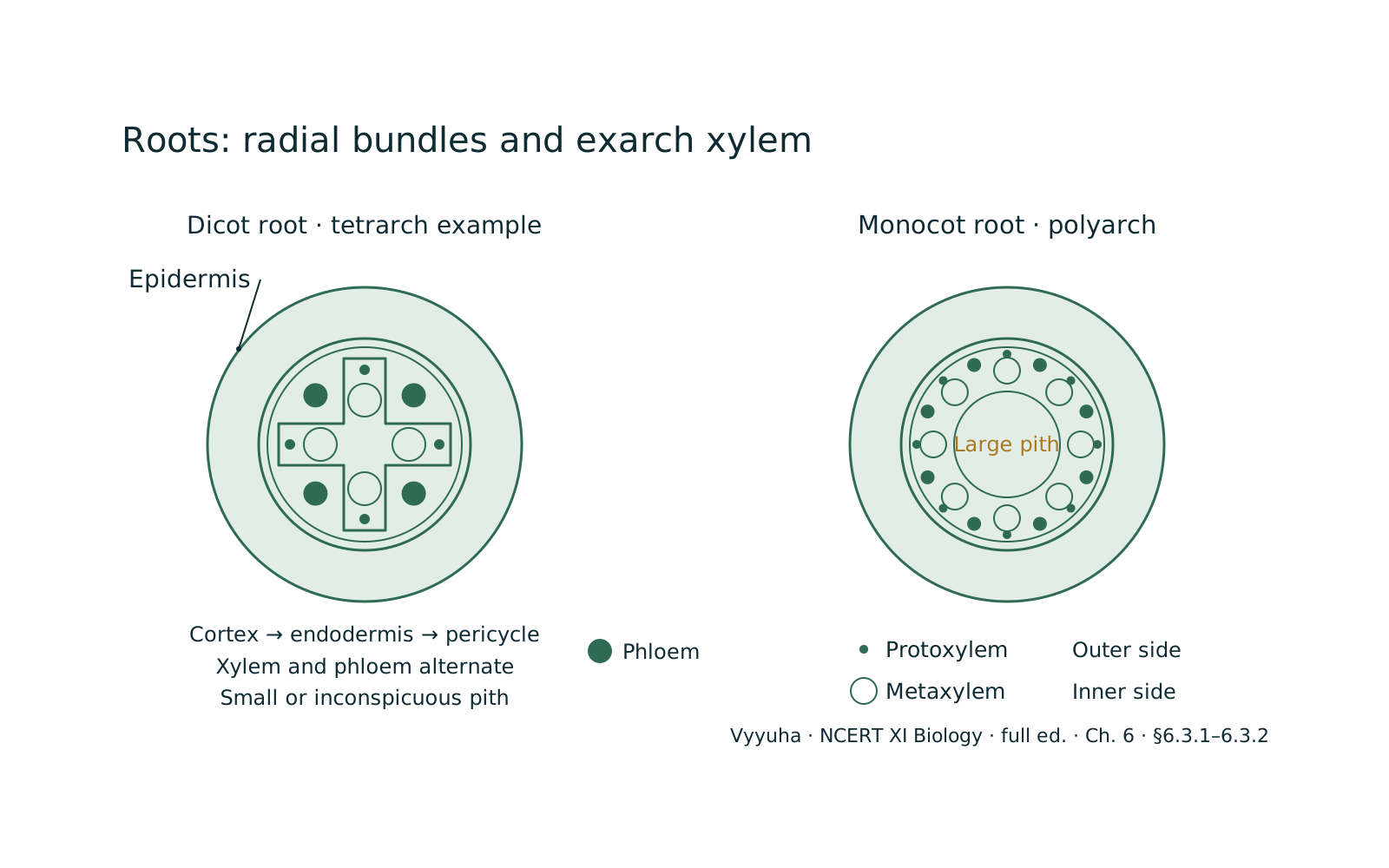
The anatomy of dicot and monocot plants reveals fundamental differences in their internal organization, crucial for understanding their biology. Dicot roots typically feature a star-shaped xylem with 2-6 arms (di- to hexarch) and a small or absent pith, while monocot roots have numerous xylem bundles (polyarch) surrounding a large central pith.
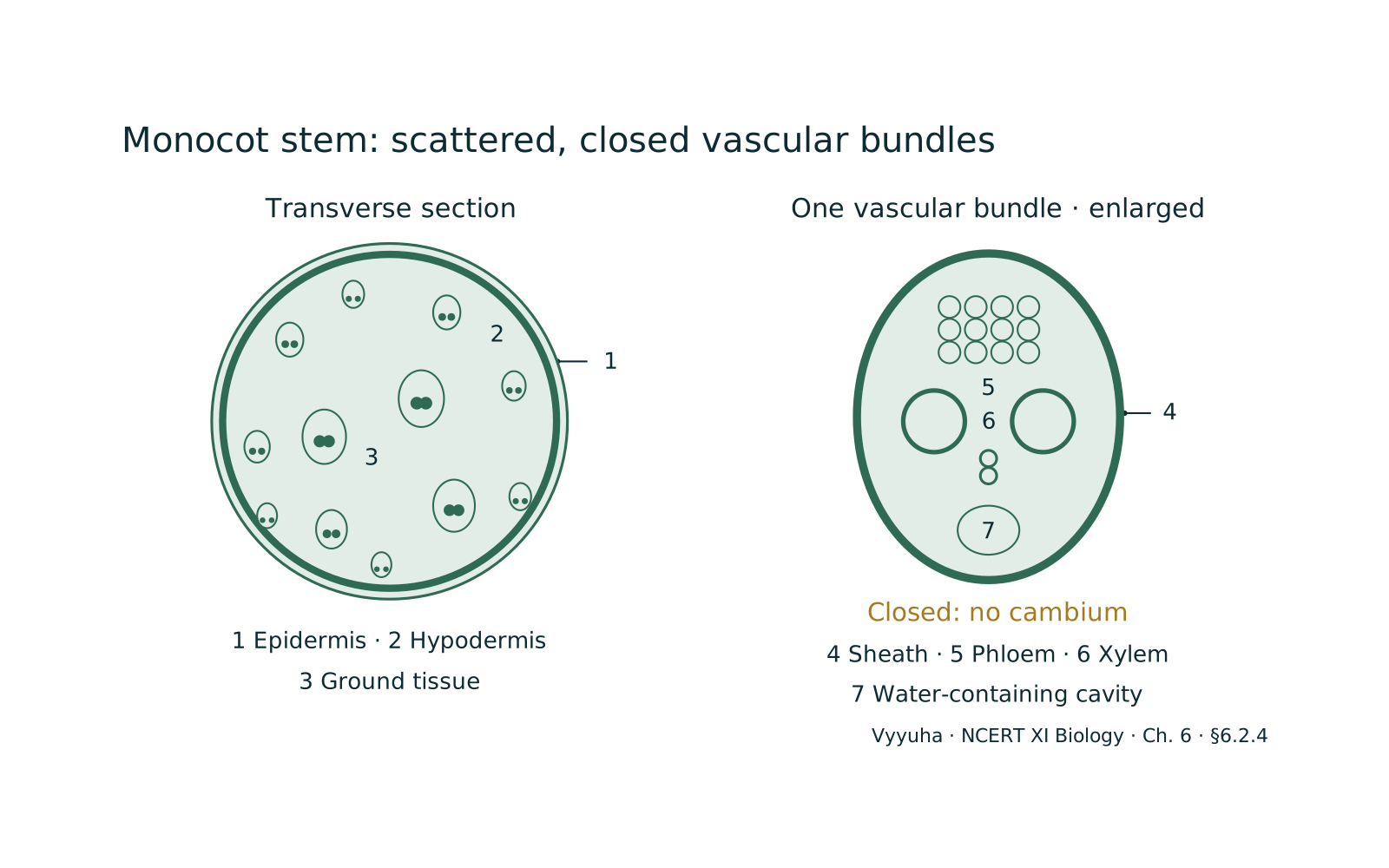
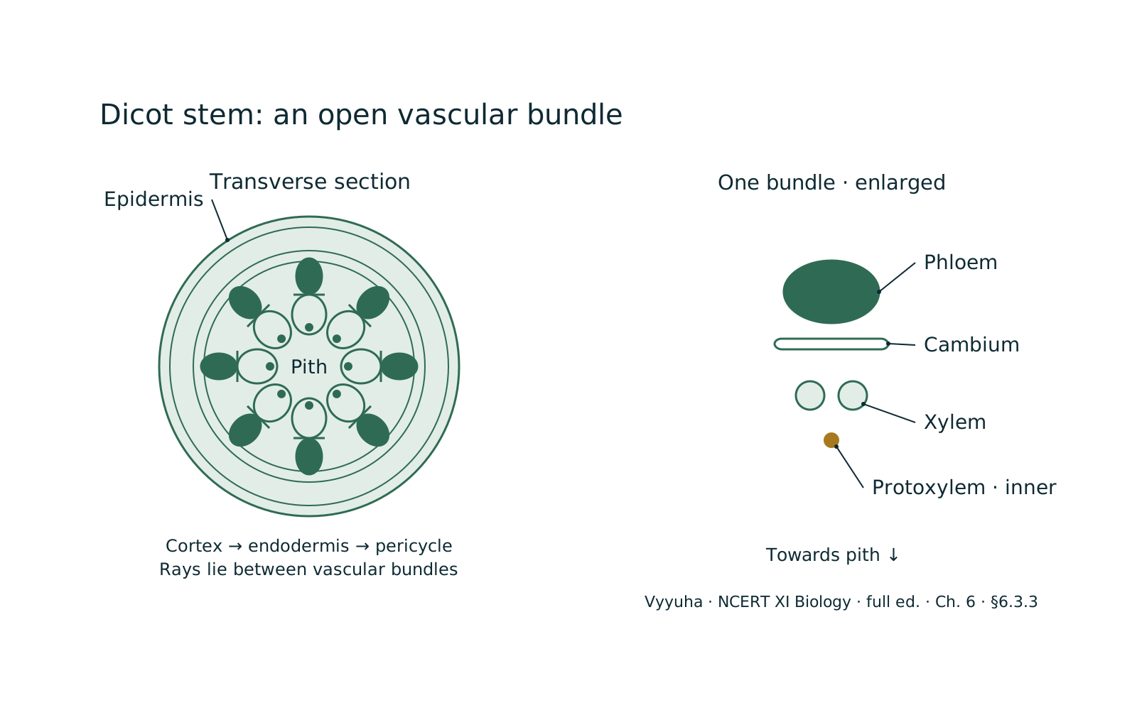
In stems, dicots exhibit vascular bundles arranged in a ring, containing cambium for secondary growth ('open' bundles), whereas monocots have scattered vascular bundles lacking cambium ('closed' bundles) and an undifferentiated ground tissue.
Dicot leaves (dorsiventral) show distinct upper and lower surfaces with differentiated mesophyll (palisade and spongy parenchyma) and reticulate venation. Monocot leaves (isobilateral) have similar upper and lower surfaces, undifferentiated mesophyll, parallel venation, and often possess bulliform cells for water conservation.
These distinctions are key diagnostic features in plant identification and reflect diverse adaptive strategies.
Full explanation
The internal organization of plant organs – roots, stems, and leaves – provides critical insights into their physiological functions and evolutionary adaptations. Dicotyledonous (dicots) and monocotyledonous (monocots) plants, while both angiosperms, exhibit distinct anatomical features that are fundamental for their classification and understanding their ecological roles.
These differences stem from their embryonic development, particularly the number of cotyledons, and manifest throughout their primary plant body.
Conceptual Foundation: The Three Tissue Systems
Before diving into specific organs, it's crucial to recall the three fundamental tissue systems that constitute the primary plant body:
- Dermal Tissue System: — The outermost protective layer, primarily composed of epidermis, which covers the entire plant body. It includes specialized cells like stomata (for gas exchange), trichomes (hairs), and root hairs (for absorption).
- Ground Tissue System: — Forms the bulk of the plant body, filling the space between the dermal and vascular tissues. It comprises parenchyma, collenchyma, and sclerenchyma cells, performing functions such as photosynthesis, storage, and support.
- Vascular Tissue System: — Responsible for long-distance transport of water, minerals, and organic nutrients. It consists of xylem (water and mineral transport) and phloem (food transport), organized into vascular bundles.
Key Principles and Evolutionary Divergence
The anatomical differences between dicots and monocots are not arbitrary but reflect distinct evolutionary pathways and adaptive strategies. For example, the presence of a vascular cambium in most dicot stems allows for secondary growth, leading to increased girth and the formation of wood, which is crucial for tall, long-lived trees.
Monocots, generally lacking a vascular cambium, typically remain herbaceous or develop alternative strategies for support (e.g., fibrous stems in palms). These structural variations are optimized for different environmental niches and growth habits.
Anatomy of Dicot Root
- Epidermis: — The outermost layer, single-layered, with numerous unicellular root hairs for water and mineral absorption. Cuticle is usually absent or very thin.
- Cortex: — A broad zone beneath the epidermis, composed of several layers of thin-walled parenchyma cells with intercellular spaces. Its primary function is food storage.
- Endodermis: — The innermost layer of the cortex, characterized by the presence of Casparian strips (suberin and lignin deposits) on its radial and tangential walls. These strips regulate the movement of water and solutes into the vascular cylinder. Some endodermal cells opposite the protoxylem are thin-walled, called passage cells, allowing water to pass.
- Stele: — The central vascular cylinder, which includes the pericycle, vascular bundles, and pith (if present).
* Pericycle: A layer of thick-walled parenchyma cells just inside the endodermis. It gives rise to lateral roots and, in dicots, contributes to the formation of vascular cambium during secondary growth.
* Vascular Bundles: Radial arrangement, meaning xylem and phloem are separate and arranged on different radii. Typically, dicot roots have 2 to 6 (diarch to hexarch) xylem bundles. The xylem is exarch (protoxylem towards the periphery, metaxylem towards the center).
* Pith: Small or absent in dicot roots. If present, it's parenchymatous and inconspicuous.
Anatomy of Monocot Root
- Epidermis: — Similar to dicot root, with unicellular root hairs.
- Cortex: — Broad, parenchymatous, with intercellular spaces, similar to dicot root.
- Endodermis: — Prominent, with Casparian strips and sometimes suberin/lignin thickening on inner walls (passage cells may be present).
- Stele:
* Pericycle: Gives rise to lateral roots. * Vascular Bundles: Radial arrangement, but typically polyarch (more than six xylem bundles, often 8 or more). Xylem is exarch. Vascular bundles are numerous. * Pith: Large, well-developed, and parenchymatous, located at the center of the stele. It often stores food.
Anatomy of Dicot Stem
- Epidermis: — Outermost protective layer, single-layered, covered by a cuticle. May bear trichomes and stomata.
- Cortex: — Located between the epidermis and the stele. It's differentiated into:
* Hypodermis: Outermost cortical layer, typically collenchymatous, providing mechanical support. * General Cortex: Parenchymatous cells with intercellular spaces, involved in storage and sometimes photosynthesis. * Endodermis (Starch Sheath): Innermost layer of the cortex, rich in starch grains, hence called the starch sheath. Casparian strips are usually absent.
- Stele: — Comprises pericycle, vascular bundles, medullary rays, and pith.
* Pericycle: Located inner to the endodermis, often a multilayered ring of sclerenchymatous or parenchymatous cells, sometimes forming a semi-lunar patch above vascular bundles. * Vascular Bundles: Conjoint (xylem and phloem together), collateral (phloem towards periphery, xylem towards center), and open (presence of cambium between xylem and phloem).
Arranged in a ring. This cambium allows for secondary growth. * Medullary Rays (Pith Rays): Radial strips of parenchymatous cells extending from the pith to the cortex, between vascular bundles. They facilitate radial conduction of water and food.
* Pith: Large, well-developed, and parenchymatous central region, primarily for storage.
Anatomy of Monocot Stem
- Epidermis: — Outermost layer, single-layered, covered by a thick cuticle. Stomata may be present.
- Hypodermis: — Sclerenchymatous, providing mechanical strength. It's usually a few layers thick.
- Ground Tissue: — Undifferentiated, meaning there's no distinct cortex, endodermis, pericycle, or pith. The entire central mass of parenchymatous cells constitutes the ground tissue, which may have intercellular spaces and store food.
- Vascular Bundles: — Conjoint, collateral, and closed (cambium is absent). They are scattered throughout the ground tissue, not arranged in a ring. Larger bundles are typically found towards the center, smaller ones towards the periphery. Each vascular bundle is usually surrounded by a sclerenchymatous bundle sheath. The xylem is Y-shaped or V-shaped, with a protoxylem lacuna (water-filled cavity) often present.
Anatomy of Dicot Leaf (Dorsiventral Leaf)
- Epidermis: — Distinct upper (adaxial) and lower (abaxial) epidermis. The upper epidermis has fewer stomata (or none), while the lower epidermis has more. Both are covered by a cuticle.
- Mesophyll: — The ground tissue between the two epidermal layers, differentiated into two types of parenchyma cells:
* Palisade Parenchyma: Elongated, vertically arranged cells, tightly packed, located below the upper epidermis. Rich in chloroplasts, primarily responsible for photosynthesis. * Spongy Parenchyma: Irregularly shaped cells with large air spaces, located below the palisade layer and extending to the lower epidermis. Facilitates gas exchange.
- Vascular Bundles: — Vary in size depending on the venation (reticulate venation). Large bundles (veins) are surrounded by bundle sheaths. Xylem is towards the upper epidermis, phloem towards the lower.
Anatomy of Monocot Leaf (Isobilateral Leaf)
- Epidermis: — Upper and lower epidermis are similar in appearance, both bearing stomata in roughly equal numbers. Covered by a cuticle. Some epidermal cells on the upper surface of grasses are large, empty, and colourless, called bulliform cells (motor cells). They help in rolling/unrolling leaves during water stress.
- Mesophyll: — Undifferentiated, meaning there's no distinction between palisade and spongy parenchyma. All mesophyll cells are roughly isodiametric and contain chloroplasts.
- Vascular Bundles: — All vascular bundles are of similar size, reflecting parallel venation, except for the main midrib vein. Each bundle is surrounded by a prominent sclerenchymatous bundle sheath. Xylem is towards the upper epidermis, phloem towards the lower.
Common Misconceptions
- All monocots have fibrous roots, all dicots have taproots: — While generally true for primary roots, many dicots (e.g., banyan) develop adventitious roots, and some monocots (e.g., maize) have a prominent primary root initially. The key difference lies in the origin and persistence of the primary root system.
- Monocots never show secondary growth: — While typical secondary growth (vascular cambium forming wood) is absent, some monocots like Dracaena and Yucca exhibit anomalous secondary growth, increasing their girth through a different mechanism.
- All leaves are dorsiventral or isobilateral: — While these are the most common types, other leaf anatomies exist, but for NEET, focusing on these two is sufficient.
NEET-Specific Angle
NEET questions frequently test the ability to distinguish between dicot and monocot organs based on their anatomical features. This often involves:
- Diagram-based questions: — Identifying a given diagram as a dicot root, monocot stem, etc.
- Feature-based questions: — Listing characteristics and asking which plant group or organ they belong to.
- Functional correlation: — Relating a specific anatomical feature (e.g., Casparian strips, bulliform cells) to its physiological role.
- Comparative analysis: — Direct comparison of features between dicots and monocots in a tabular format or multiple-choice options. Mastery of the distinguishing features is paramount for scoring well on this topic.
Key Concepts
Vascular bundles are the plant's transport highways. In dicot stems, they are 'open' (meaning they have a…
The stele is the central core of the root, containing the vascular tissues. In dicot roots, the xylem forms a…
The mesophyll is the photosynthetic tissue of the leaf. In dicot leaves (dorsiventral leaves), the mesophyll…
Often confused with
Side-by-side differences the NEET paper likes to test.
| Aspect | Anatomy of Dicot and Monocot Plants | Monocot Plants |
|---|---|---|
| Root Xylem Bundles | Dicot Root: 2-6 (diarch to hexarch), star-shaped | Monocot Root: More than 6 (polyarch), often in a ring around pith |
| Root Pith | Dicot Root: Small or absent | Monocot Root: Large and well-developed |
| Stem Vascular Bundles Arrangement | Dicot Stem: Arranged in a ring | Monocot Stem: Scattered throughout ground tissue |
| Stem Vascular Bundles Type | Dicot Stem: Open (cambium present), conjoint, collateral | Monocot Stem: Closed (cambium absent), conjoint, collateral |
| Stem Ground Tissue | Dicot Stem: Differentiated into cortex, endodermis, pericycle, pith | Monocot Stem: Undifferentiated ground tissue |
| Leaf Mesophyll | Dicot Leaf (Dorsiventral): Differentiated into palisade and spongy parenchyma | Monocot Leaf (Isobilateral): Undifferentiated |
| Leaf Bulliform Cells | Dicot Leaf: Absent | Monocot Leaf: Often present on upper epidermis |
| Leaf Venation | Dicot Leaf: Reticulate (net-like) | Monocot Leaf: Parallel |
| Secondary Growth | Dicot: Typically present in stems and roots | Monocot: Generally absent (some anomalous exceptions) |
The anatomical distinctions between dicot and monocot plants are pervasive across their roots, stems, and leaves, reflecting their divergent evolutionary paths and adaptive strategies. Key differences include the number and arrangement of vascular bundles in roots and stems, the presence or absence of a central pith, the differentiation of ground tissues, and the internal organization of leaf mesophyll.
These features, such as the open vascular bundles in dicot stems enabling secondary growth versus closed bundles in monocots, or the presence of bulliform cells in monocot leaves, are crucial for their survival and provide reliable diagnostic characteristics for identification.
Why it is tested: For NEET, understanding these anatomical differences is critical for identifying plant types from diagrams, correlating structure with function (e.g., Casparian strips, bulliform cells), and answering comparative questions. It forms a foundational understanding for plant physiology and ecology.
Questions students ask
5 answered on this topic.
What is the primary difference in vascular bundle arrangement between dicot and monocot stems?
In dicot stems, vascular bundles are typically arranged in a distinct ring around a central pith. These bundles are 'open' because they contain a vascular cambium between the xylem and phloem, allowing for secondary growth. In contrast, monocot stems have vascular bundles scattered throughout the ground tissue, without a clear ring arrangement. These bundles are 'closed' as they lack a vascular cambium, which generally prevents secondary growth in thickness.
How do Casparian strips in the endodermis function in roots?
Casparian strips are bands of suberin and lignin found on the radial and tangential walls of endodermal cells in roots. Their primary function is to block the apoplastic pathway (movement through cell walls and intercellular spaces) for water and dissolved solutes. This forces water and minerals to enter the symplastic pathway (through the cytoplasm and plasmodesmata) of the endodermal cells, allowing the plant to selectively regulate what enters the vascular cylinder (stele).
What are bulliform cells and what is their role in monocot leaves?
Bulliform cells, also known as motor cells, are large, empty, and colourless epidermal cells found on the upper surface of many monocot leaves, particularly grasses. They are specialized for water storage. When turgid, they help keep the leaf expanded. During water stress, they lose turgor, causing the leaf to roll inwards, thereby reducing the exposed surface area and minimizing water loss through transpiration.
Why do dicot stems typically show secondary growth, while monocot stems generally do not?
Dicot stems possess a vascular cambium, a lateral meristem located between the xylem and phloem within their vascular bundles (making them 'open'). This cambium actively divides to produce secondary xylem inwards and secondary phloem outwards, leading to an increase in stem girth, known as secondary growth. Monocot stems, however, lack this vascular cambium in their vascular bundles (making them 'closed'), which is why they generally do not undergo typical secondary growth.
Explain the difference between dorsiventral and isobilateral leaves.
Dorsiventral leaves, typical of dicots, have distinct upper (dorsal) and lower (ventral) surfaces. Internally, their mesophyll is differentiated into palisade parenchyma (densely packed, columnar cells below the upper epidermis) and spongy parenchyma (irregularly shaped cells with large air spaces near the lower epidermis).
Isobilateral leaves, characteristic of monocots, have similar upper and lower surfaces, and their mesophyll is undifferentiated, meaning it consists of a single type of parenchymatous cells without distinct palisade and spongy layers.
Stomata are also more evenly distributed on both surfaces in isobilateral leaves.
Revise in 30 seconds
- Dicot Root: — Di-hexarch xylem, small/absent pith, radial bundles.
- Monocot Root: — Polyarch xylem, large pith, radial bundles.
- Dicot Stem: — Vascular bundles in a ring, open (cambium present), collenchymatous hypodermis, differentiated ground tissue.
- Monocot Stem: — Vascular bundles scattered, closed (cambium absent), sclerenchymatous hypodermis, undifferentiated ground tissue.
- Dicot Leaf (Dorsiventral): — Differentiated mesophyll (palisade & spongy), reticulate venation, more stomata on lower epidermis.
- Monocot Leaf (Isobilateral): — Undifferentiated mesophyll, parallel venation, bulliform cells, stomata on both surfaces.
For Dicot vs. Monocot: 'R.S.L. - D.M.':
Root:
- Dicot: Di-hexarch, Diminutive Pith (Small/Absent)
- Monocot: Many Xylem (Polyarch), Massive Pith (Large)
Stem:
- Dicot: Distinct Ring, Differentiated Ground, Does Secondary Growth (Open Bundles)
- Monocot: Mixed/Scattered, Monotonous Ground (Undifferentiated), Missing Secondary Growth (Closed Bundles)
Leaf:
- Dicot: Dorsiventral, Differentiated Mesophyll, Diverse Veins (Reticulate)
- Monocot: Motor Cells (Bulliform), Monotonous Mesophyll (Undifferentiated), Many Parallel Veins