Proteins — Explained
Detailed Explanation
Proteins are arguably the most diverse and functionally significant class of macromolecules in living systems. Their unparalleled versatility stems from the combinatorial possibilities offered by 20 standard amino acids and the intricate folding patterns they adopt. To truly grasp the essence of proteins, one must delve into their fundamental building blocks, the bonds that link them, and the hierarchical levels of their structural organization.
Conceptual Foundation: The Amino Acid Building Blocks
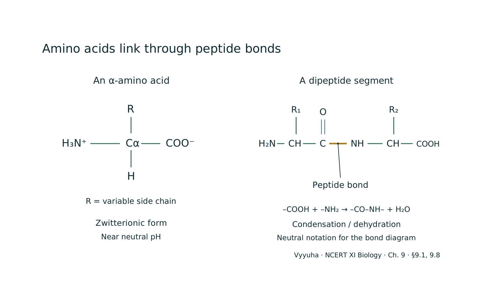
At the heart of every protein lies the amino acid. Each amino acid possesses a central carbon atom, known as the alpha-carbon (-carbon), to which four different groups are attached: an amino group (), a carboxyl group (), a hydrogen atom (), and a unique side chain ( group).
It is this group that distinguishes one amino acid from another and confers its specific chemical properties – whether it's polar, nonpolar, acidic, basic, or contains sulfur. The presence of both an amino and a carboxyl group allows amino acids to act as zwitterions, meaning they can exist in a dipolar ionic form where the amino group is protonated () and the carboxyl group is deprotonated () at physiological pH.
Amino acids are broadly classified based on the nature of their R-groups:
- Nonpolar, Aliphatic R-groups: — Glycine, Alanine, Valine, Leucine, Isoleucine, Methionine, Proline.
- Aromatic R-groups: — Phenylalanine, Tyrosine, Tryptophan.
- Polar, Uncharged R-groups: — Serine, Threonine, Cysteine, Asparagine, Glutamine.
- Positively Charged (Basic) R-groups: — Lysine, Arginine, Histidine.
- Negatively Charged (Acidic) R-groups: — Aspartate, Glutamate.
Of the 20 common amino acids, some are termed 'essential' because the human body cannot synthesize them and they must be obtained from the diet (e.g., Leucine, Isoleucine, Valine, Lysine, Methionine, Phenylalanine, Threonine, Tryptophan, Histidine). Others are 'non-essential' as the body can synthesize them. This distinction is crucial for understanding nutritional requirements.
Peptide Bond Formation: Linking Amino Acids
Amino acids link together to form long chains through a special type of covalent bond called a peptide bond. This bond forms between the carboxyl group of one amino acid and the amino group of another, with the elimination of a water molecule (a dehydration reaction).
The resulting bond is rigid and planar, possessing partial double-bond character due to resonance. A chain of amino acids linked by peptide bonds is called a polypeptide. By convention, polypeptides are written from the N-terminus (free amino group) to the C-terminus (free carboxyl group).
Key Principles: Levels of Protein Structure
The functional diversity of proteins arises from their ability to fold into precise three-dimensional structures. This folding process is hierarchical, described by four levels of organization:
- Primary Structure: — This is the simplest level, referring to the unique linear sequence of amino acids in a polypeptide chain. It is determined by the genetic code and is crucial because it dictates all subsequent levels of structure. Even a single amino acid change can drastically alter a protein's function (e.g., sickle cell anemia, where a single glutamate is replaced by valine in hemoglobin).
- Secondary Structure: — Localized, regularly repeating structures formed by hydrogen bonding between the backbone atoms (not R-groups) of the polypeptide chain. The two most common types are:
* **Alpha-helix (-helix):** A right-handed coiled structure where each carbonyl oxygen forms a hydrogen bond with the amide hydrogen of an amino acid four residues ahead ( to ). The R-groups project outwards from the helix.
* **Beta-pleated sheet (-pleated sheet):** Consists of two or more polypeptide strands (beta strands) arranged side-by-side, connected by hydrogen bonds between backbone atoms. The strands can be parallel (N-termini aligned) or antiparallel (N-terminus of one aligned with C-terminus of another).
R-groups project above and below the plane of the sheet. * Supersecondary Structures (Motifs): Specific combinations of secondary structures that appear in various proteins, e.g., motif, helix-loop-helix.
- Tertiary Structure: — The overall three-dimensional shape of a single polypeptide chain, including the spatial arrangement of all its atoms. It results from interactions between the R-groups of amino acids, as well as interactions between R-groups and the polypeptide backbone. These interactions include:
* Hydrophobic interactions: Nonpolar R-groups tend to cluster in the interior of the protein, away from water. * Ionic bonds (salt bridges): Electrostatic interactions between oppositely charged R-groups (acidic and basic amino acids).
* Hydrogen bonds: Between polar R-groups, or between polar R-groups and backbone atoms. * Disulfide bonds: Covalent bonds formed between the sulfhydryl groups () of two cysteine residues, creating a strong cross-link that stabilizes the tertiary structure.
These are particularly important in extracellular proteins. * Van der Waals forces: Weak, transient attractive forces between all atoms. The tertiary structure often contains distinct functional units called 'domains', which are independently folding regions within a polypeptide.
- Quaternary Structure: — This level applies only to proteins composed of two or more polypeptide chains (subunits), which are held together by non-covalent interactions (and sometimes disulfide bonds). The arrangement of these subunits in space constitutes the quaternary structure. Examples include hemoglobin (four subunits), antibodies (four subunits), and many enzymes. Each subunit typically has its own primary, secondary, and tertiary structure.
Protein Folding and Denaturation
Protein folding is a highly complex and spontaneous process, guided by the primary sequence, where a polypeptide chain acquires its specific 3D functional structure. This process is often assisted by 'chaperone proteins' (heat shock proteins) that prevent misfolding or aggregation. Misfolding can lead to serious diseases like Alzheimer's, Parkinson's, and Creutzfeldt-Jakob disease (prion diseases).
Denaturation is the process by which a protein loses its native, functional three-dimensional structure due to the disruption of its secondary, tertiary, and quaternary structures. The primary structure (peptide bonds) usually remains intact.
Denaturation can be caused by physical agents (heat, radiation, mechanical agitation) or chemical agents (strong acids/bases, organic solvents, heavy metal ions, detergents). While some proteins can undergo renaturation (regain their structure and function) if the denaturing agent is removed, many denatured proteins are irreversibly altered.
Real-World Applications and Functions
Proteins are involved in virtually every biological process:
- Enzymatic Catalysis: — Enzymes are highly specific protein catalysts that accelerate biochemical reactions (e.g., amylase, pepsin, DNA polymerase).
- Structural Support: — Provide strength and rigidity to cells and tissues (e.g., collagen in connective tissue, keratin in hair/nails, actin/myosin in muscle).
- Transport: — Carry specific molecules across membranes or throughout the body (e.g., hemoglobin for oxygen, albumin for fatty acids, membrane channels/pumps).
- Immune Defense: — Antibodies (immunoglobulins) recognize and neutralize foreign invaders.
- Movement: — Motor proteins facilitate cellular and organismal movement (e.g., actin and myosin in muscle contraction, dynein and kinesin in intracellular transport).
- Signaling and Regulation: — Hormones (e.g., insulin, growth hormone) and receptors transmit signals, regulating cellular processes.
- Storage: — Store essential nutrients (e.g., ferritin stores iron, ovalbumin stores amino acids in egg white).
Common Misconceptions
- All proteins are enzymes: — While many proteins are enzymes, not all proteins function as catalysts. Many have structural, transport, or regulatory roles.
- All enzymes are proteins: — This is largely true, but there are exceptions. Ribozymes, which are RNA molecules, can also exhibit catalytic activity.
- Denaturation always breaks peptide bonds: — Denaturation primarily disrupts the weaker non-covalent interactions (hydrogen bonds, ionic bonds, hydrophobic interactions) and disulfide bonds that maintain secondary, tertiary, and quaternary structures. The strong covalent peptide bonds of the primary structure are generally unaffected unless extreme conditions are applied (e.g., strong acid hydrolysis).
NEET-Specific Angle
For NEET, a strong emphasis is placed on:
- Amino acid classification and essentiality: — Memorizing examples of essential amino acids and understanding the properties of different R-groups.
- Peptide bond formation: — Understanding the dehydration reaction and its characteristics.
- Levels of protein structure: — Being able to identify and differentiate between primary, secondary (-helix, -sheet), tertiary, and quaternary structures, along with the types of bonds/interactions stabilizing each level. Specific examples like hemoglobin for quaternary structure are important.
- Protein functions: — Associating specific proteins with their roles (e.g., collagen-structural, hemoglobin-transport, antibodies-defense, enzymes-catalysis).
- Denaturation: — Understanding its causes and consequences, and the concept of renaturation.
- Diseases related to protein structure/function: — Sickle cell anemia (point mutation affecting primary structure), prion diseases (misfolding), enzyme deficiencies.
- Chaperone proteins: — Their role in proper protein folding.
Mastering these aspects will provide a solid foundation for tackling NEET questions related to proteins.
Often confused with
Side-by-side differences the NEET paper likes to test.
| Aspect | Proteins | Carbohydrates |
|---|---|---|
| Monomer Units | Amino acids (20 common types) | Monosaccharides (e.g., glucose, fructose, galactose) |
| Bond Type | Peptide bonds | Glycosidic bonds |
| Primary Function | Diverse: enzymes, structural, transport, defense, signaling, movement | Primary energy source, structural components (e.g., cellulose, chitin), cell recognition |
| Elemental Composition | C, H, O, N (often S) | C, H, O (ratio $1:2:1$ typically) |
| Structural Complexity | Highly complex 3D structures (primary to quaternary) | Linear or branched chains, less complex 3D folding compared to proteins |
Proteins and carbohydrates are both essential biomolecules, but they differ fundamentally in their building blocks, bonding, and primary roles. Proteins are polymers of amino acids linked by peptide bonds, characterized by their nitrogen content and highly diverse, intricate three-dimensional structures that enable a vast array of functions.
Carbohydrates, on the other hand, are polymers of monosaccharides joined by glycosidic bonds, primarily serving as energy sources and structural components, and typically adhering to a carbon:hydrogen:oxygen ratio of 1:2:1.
Their distinct chemical compositions and structural complexities underpin their specialized roles in biological systems.
Why it is tested: NEET relevance: Understanding the fundamental differences between proteins and carbohydrates is crucial for NEET aspirants. Questions often test the identification of monomers, types of bonds, and primary functions of these macromolecules. For instance, distinguishing between peptide bonds and glycosidic bonds, or identifying the presence of nitrogen as a key characteristic of proteins (unlike pure carbohydrates), are common conceptual checks. Knowledge of their diverse functions helps in relating structure to biological roles, which is a recurring theme in NEET Biology.
Questions students ask
5 answered on this topic.
What makes one amino acid different from another?
The distinguishing feature among the 20 common amino acids is their unique side chain, also known as the R-group. While all amino acids share a common backbone structure (an alpha-carbon, an amino group, a carboxyl group, and a hydrogen atom), the R-group varies greatly in its chemical composition, size, shape, charge, and polarity.
This diversity in R-groups is what confers the specific properties to each amino acid, influencing how it interacts with other amino acids and its environment, ultimately determining the protein's overall structure and function.
Can proteins regain their original shape after denaturation?
Yes, some proteins can regain their original functional shape, a process known as renaturation, if the denaturing agent is removed and the conditions are returned to normal. This phenomenon demonstrates that the primary sequence of amino acids contains all the necessary information for a protein to fold into its correct three-dimensional structure.
However, many proteins, especially those that have undergone severe or prolonged denaturation, cannot renature and remain irreversibly denatured, losing their biological activity permanently. The ability to renature depends on the specific protein and the extent of denaturation.
What is the significance of disulfide bonds in protein structure?
Disulfide bonds are strong covalent bonds formed between the sulfhydryl (-SH) groups of two cysteine residues. They play a crucial role in stabilizing the tertiary and sometimes quaternary structures of proteins, particularly those found in extracellular environments where they provide significant structural rigidity and resistance to denaturation.
These bonds act as molecular staples, locking specific regions of the polypeptide chain into place, which is vital for maintaining the protein's functional conformation and overall stability. They are less common in intracellular proteins due to the reducing environment within cells.
How do chaperone proteins assist in protein folding?
Chaperone proteins, often referred to as heat shock proteins (HSPs), are a class of proteins that assist in the proper folding of other proteins. They do not dictate the final folded structure but rather prevent incorrect folding pathways and aggregation of nascent or partially folded polypeptides.
Chaperones bind to hydrophobic regions of unfolded proteins, shielding them from the aqueous environment and preventing premature interactions that could lead to misfolding or aggregation. They provide a protected environment for proteins to fold correctly, often utilizing ATP hydrolysis to facilitate conformational changes that aid in the folding process, ensuring cellular proteostasis.
What are essential amino acids and why are they important?
Essential amino acids are those amino acids that the human body cannot synthesize on its own or cannot synthesize in sufficient quantities to meet its metabolic needs. Therefore, they must be obtained through the diet.
There are nine essential amino acids for adults: histidine, isoleucine, leucine, lysine, methionine, phenylalanine, threonine, tryptophan, and valine. They are critically important because they are the fundamental building blocks for all proteins synthesized in the body.
A deficiency in even one essential amino acid can impair protein synthesis and lead to various health issues, highlighting the importance of a balanced diet rich in complete proteins.